IDIOPATHIC SCOLIOSIS |
X-RAY EVALUATION
It must be carried out making X-rays of the backbone from the occiput to the sacrum, standing straight, in the two antero-posterior and lateral projections (30 x 80 X-ray photographs).
The X-rays can reveal:
-
the seat of the curves
- the degrees of the curves according to Cobb’s method, which can be easily used sticking to the following rules (fig. 12): first of all, you must trace out two horizontal lines of which one on the upper edge of the upper terminal vertebra and the other on the lower edge of the lower terminal vertebra (the terminal vertebrae are the ones situated no the upper and lower limits of a curve, and whose maximum inclination is situated towards the concavity of the curve); then you must race out two more lines perpendicularly to the previous ones, measuring their angle of intersection. Cobb’s method, rather than Risser-Ferguson’s, is recommended by the Scoliosis Research Society. The broken arrows in the figure do not converge towards the concavity to be measured, so that the vertebrae are not terminal and are in another curve situated above and below the curve to be measured.
-
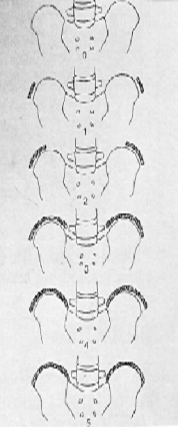
The patient’s actual or physiological osseous age (which does not always coincide with the chronological age) is evaluated through the Risser’s test, which studies the maturation stage of the ossification core of the iliac wing (fig. 13) or though the research of the epiphyseal ossification cores of the vertebra (the so-called “ring apophysis”) (fig. 14).
- The possible presence of congenital malformations with particular attention to the lumbosacral passage (spondylolysis and spondylolisthesis).
.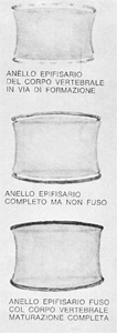
fig 14 – Direct evaluation of
the growth degree of the
backbone through the
presence of the
vertebral “ring apophysis”
|
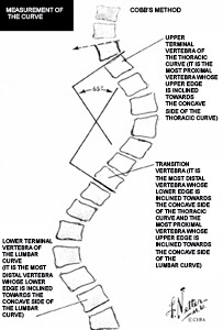
Fig 12 - See description in the text
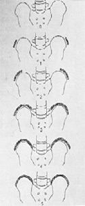
Fig. 13 -
Risser 0 = there is no ossification core on the iliac crest;
Risser 1+ = initial presence of the core with a covering of 1/4 of the iliac crest;
Risser 2+ = covering of 2/4 of the iliac crest;
Risser 3+ = covering of 3/4 of the iliac crest;
Risser 4+ = covering of 4/4 of the iliac crest;
Risser 5+ = fusion occurred; the backbone has by now stabilized the growth.
Risser’s sign is an indirect evaluation of the growth degree of the backbone.
|
|
|


